Reproductive system of Herdmania:
Herdmania is bisexual or hermaphrodite. Self fertilization does not occur due to protogynous condition.
(A) GONADS
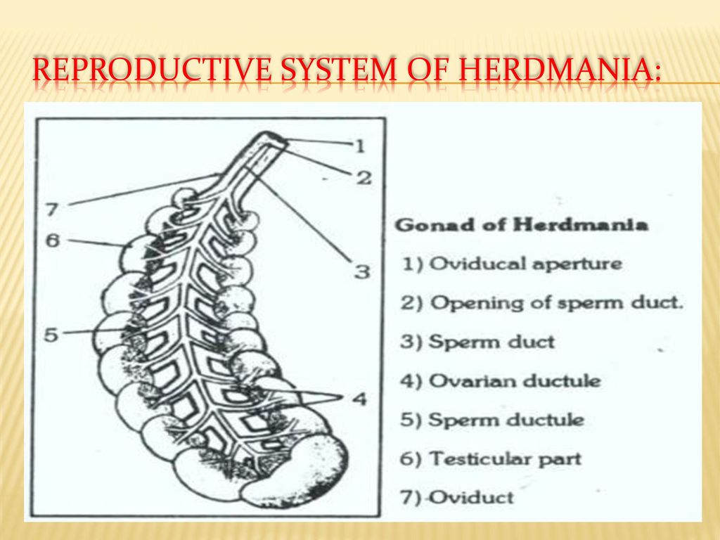
There is present a pair of gonads embedded in mantle on either side. The right gonad lies above the pericardium and left in intestinal loop. Each gonad is an elongated structure with 10-25 distinct rounded lobes arranged in two rows on either side of a central axis (Fig. 18). The median lobe present at proximal end is the largest, single and bean shaped. Each lobe has peripheral, large, cortical, brick red coloured testicular region and inner, smaller, medullary, pinkish coloured ovarian region. The testicular region caeca lined by germinal epithelium forming spermatogonia, spermatocytes and spermatozoa. The ovarian region ns of siphons consists of ovarian follicles containing rounded ova in various stages of development. So each lobe of gonad is a hermaphrodite gland.
(B) GONODUCTS: Each gonad has an oviduct and a ves deferens ,both running parallel along the central axis. The vas deferens or spermatic duct is a narrower duct formed by the union of spermatic ductules coming from testicular zone of each lobe.The oviduct is wider tube formed by the joining of ovarian ductules coming from ovarian zoneof each lobe. The gonoducts from both sides extend beyond gonads and open independently into the cloaca.
(C) GAMETES
Both type of gametes are produced by gonads. These are carried through gonoducts into cloaca and then outside with outgoing water current.
(i) Sperms : These are very minute about 4 mm in length. Each sperm has anterior broad head having nucleus and beakshaped acrosome, a short middle piece and a very straight long tail. The sperms show polymorphism on the basis of size of acrosome.
(ii) Ova. These are formed in ovarian part of gonad. A mature ovum is about 3 mm in diameter It contains small amount of yolk (microlecithal). It has a large nucleus present on one side with a large nucleolus and dense chromatin granules. The ovum is surrounded by three membranes: (a) Vitelline membrane, (b) inner chorion and (c) outer chorion. The space between vitelline membrane and inner chorion, and inner chorionand interchorionic fluid. and outer chorion is filled with perivitelline fluid