Reproduction of Selaginella:
VEGETATIVE REPRODUCTION : It takes place by following methods:
1. Fragmentation: Under humid conditions, plants develop adventitious roots and branches which on separation from the parent plant, due to death decay or mechanical forces like wind or air etc., grow into independent plants.
2. Tubers: At the end of growing season some species and bear tubers at the tip of special narrow underground branches arising from the base of stem. Tubers store food and bear rudimentary scales. During unfavorable conditions when the aerial parts die, the tubers survive and perennate and on the advent of favourable conditions, they grow into new plants.
3. Resting buds: At the end of growing season, some sp. develop resting buds at the apices of aerial vegetative branches. The leaves are closely appressed and cover the growing point in this region. These buds survive under the adverse conditions when the whole plant dies. On return of favorable conditions these develop into new plants by producing rhizophores that bear roots to fix them to the substratum.
REPRODUCTION BY SPORES
All the species of Selaginella are heterosporous ie it reproduces asexually by means of two types of spores The smaller microspores and larger megaspores produced by meiosis in their respective sporangium. Haploid spores germinate to produce male and female gametophytes.
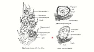
THE STROBILUS OR CONE.
The spores are produced in side the sporangia borne in the axils of leaves called sporophylls arranged in definite loose or compact terminal structures called cones or strobilus or spike.
Strobili are borne on the terminal ends of main axis or lateral branches and with their formation, apical growth of the branches stop. It is 0.5 to 5 cm in length, cylindrical or dorsiventral in shape. It consists of central axis on which the sporophytes are spirally arranged and are isophyllous. The sporophyll bearing microsporangia are called microsporophylls and that bearing megasporangia are called megasporophylls. There is a great variation in the distribution of micro and megasporophylls on the strobilus.
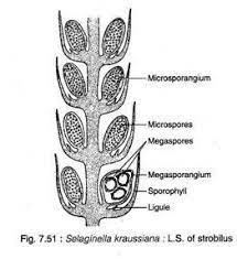
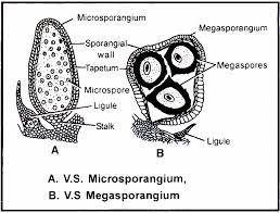
THE SPORANGIUM
1. Structure of Microsporangium : Each microsporangium is reddish brown in oval, rein form or spherical body attached in the axils of microsporophylls in between the ligule and axis with the help of a short multicellular stalk. It has a two-layered jacket wall consisting of thick walled columnar cells containing chloroplast when young. At maturity radial and inner tangential wall of cells of outer layer get thickened whereas cells of inner layer remain thin walled. Next to the inner layer is the tapetal layer, which is nutritive in function and encloses a large number of microspore mother cells. The latter undergo meiosis to produce numerous microspore tetrads, which later on get separate. Thus at maturity cavity of microsporangium is filled with about 1500-2000 haploid microspores surrounded by only single layered jacket as tapetum and inner wall disintegrate.
2. Structure of Megasporangium : It is comparatively large, green cream or dark brown in color, four lobed structure attached in the axils ofmegasporophylls between the ligule and axis with the help of a short multicellular stalk. It has two layered Jacket wall followed by a tapetal layer enclosing a number of megaspore mother cells. Out of which only one remain functional and divides meiotically to form tetrads of four haploid megaspores. By this time outer megasporangial wall became thick and radially elongated and inner wall and tapetum disintegrates so that megaspores get separate and grow considerably in size. Due to abortion of megaspore number may varies in size.
MALE GAMETOPHYTE
1. MICROSPORE:
Microspores are much smaller ranging from .015 to 05 mm in diameter and tetrahedral in shape with rounded base and distinct tri-radiate mark. Each spore possess a single nucleus surrounded by cytoplasm and enclosed by two layered spore wall. The outer, thick and variously sculptured is exine or exospore and inner delicate and thin is intine or endospore. The exine contains sporopollenin (a waxy substance) and carbohydrates, the intine contains cellulose and polysaccharides and the cytoplasm contains reserve food in the form of oil
globules and fatty substance.
GERMINATION:
Germination of microspore is precocious i.e. It starts germination within the microsporangium. At 13-celled stage the micro gametophytes are shed from the microsporangium and further development takes place on the substratum. By repeated mitotic division, 128-216 androcytes (mother cells) are produced, each of which metamorphose into antherozoids.
Each spermatozoid is an elongated narrow biflagellate structure measuring about 25 micrometer in length. One flagellum is smaller and attached to the extreme anterior and the other largest is a little below the former. The nucleus encloses the posterior half enclosed within the cytoplasm.
FEMALE GAMETOPHYTE
MEGASPORE :
Each megaspore is a large, whitish or light brown in color and tetrahedral in shape with a rounded base and distinct tri-radiate mark. Each spore possess a nucleus surrounded by cytoplasm rich in oils and fatty substances and enclosed by a two-layered thick wall envelope. The outer layer is thick and variously sculptured called the exine or exospores, which is further differentiated into an outer heterogeneous ectine and inner homogenous endine or hexine. The inner layer is thin and hyaline called intine or endospore.
GERMINATION:
The megaspores start germinating while they are still enclosed with in the megasporangia i.e. In situ, and are shed at various stages of development. The germination of megaspore is precocious. It obtains nourishment from the tapetal fluid and start germination. At first endine separates from exine due to greater growth of latter, creating a fluid filled space between the two.