Digestive system of Herdmania:
The digestive system of herdmania is divided into two parts-
(1) Alimentary canal
(2) Digestive gland
(1) Alimentari canal:The alimentary canal is complete,coiled and u shaped. It has following parts:
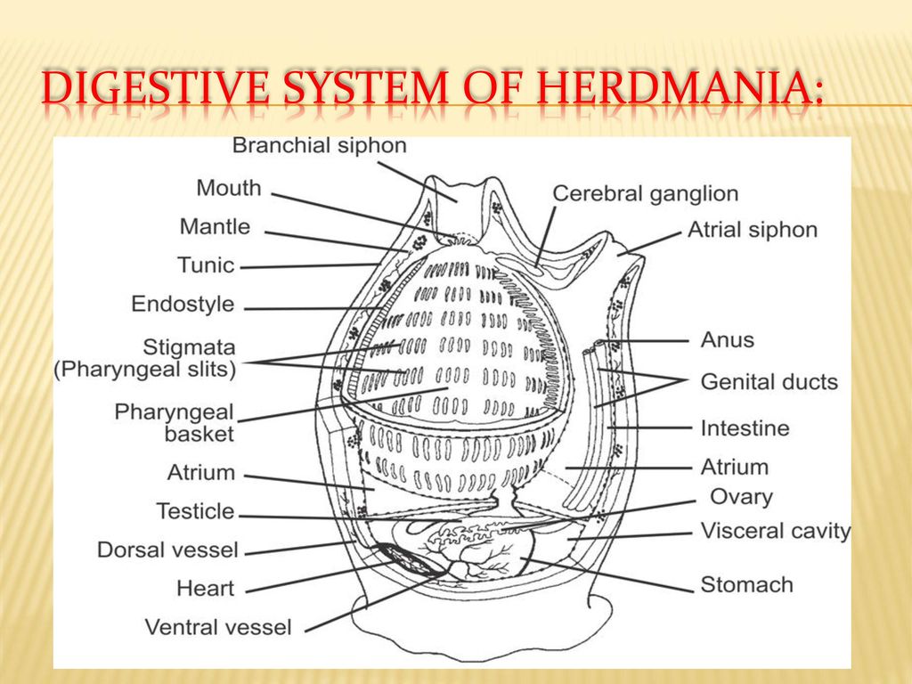
(i) Mouth. It is present at tip of the branchial siphon and also called branchial aperture. It is bordered by four lips. It leads into buccal cavity.
(ii) Buccal cavity. It is lined by epidermis, so called as stomodaeum. It is short, narrow and laterally compressed cavity. It has at its base a branchial sphincter to regulate its opening. Below the sphincter, there are present highly branched delicate branchial tentacles about 64 in number. The free ends of tentacles meet at the centre to act as stainer. So tentacles allow entry of the microscopic food particles into the pharynx.
(iii) Pharynx Buccal cavity opens into pharynx. It is largest part of alimentary canal. It is divided into pre branchial zone and a branchial sac.
(a) Pre branchial zone. It is a smaller anterior region of pharynx demarcated from the branchial sac by anterior and posterior peripharyngeal bands. In between these two bands peripharyngeal groove is present. The large cilia are present on bands whereas short cilia are present in groove. The anterior peripharyngeal band is a complete ring whereas posterior peripharyngeal band is interrupted mid dorsally by the dorsal lamina and mid ventrally by the endostyle. Dorsal tubercle is present in front of dorsal lamina.
(b) Branchial sac. It is a large basket like posterior part of pharynx. It is also called branchial basket. Its lateral walls are perforated by numerous gill slits or stigmata. Each side of branchial sac has about 200,000 stigmata. The stigmata bear long cilia on margins called lateral cilia. The lateral walls of branchial sac has network of broad longitudinal and transverse bars at regular intervals enclosing many squarish or rectangular areas called stigmatic areas (Fig. 7). Each stig- matic area has 5 or 6 stigmata. In stigmatic area, interstigmatic bars are present between adjacent stigmata.
The outer wall of branchial sac is connected to mantle by hollow strands called trabeculae. The inner wall has 8-10 branchial folds on either side. A thin flap or fold is suspended from short mid dorsal border of roof of branchial sac called dorsal lamina or hyperpharyngeal band. It runs from posterior peripharyngeal band to oesophagus. It has 20-30 conical tapering tongue like processes hanging into branchial sac called languets.
Similarly, along anterior and ventral border of branchial sac is present a mid ventral groove called endostyle. It is continuous with peripharyngeal groove anteriorly. Posteriorly, the groove ends before oesophagus and extends upto oesophagus as retropharyngeal bands. The endostyle is lined by five longitudinal ciliated tracts (1 median, 2 lateral pairs) alternating with four longitudinal tracts of mucus secreting cells.
The posteriormost part of pharynx has a small circular area made of two semicircular lips guarding oesophageal opening called oesophageal area. It lacks stigmata and blood vessels.
(iv) Oesophagus. It is a very short, curved, thick-walled tube opening into the stomach. It has four longitudinal ciliated grooves to direct food into the stomach.
(v) Stomach. It is a thin-walled tube, wider than oesophagus. It has sphincters at both ends. It is surronded on either side by right and left lobe of liver.
(vi) Intestine. It is a thin walled, U-shaped tube having a proximal descending limb and a distal ascending limb. The intestinal loop encloses the left gonad. It leads into rectum.
(vii) Rectum. It is a short, narrow tube opening dorsally into atrium or cloaca through anus. It is internally lined by cilia.
(viii) Anus. It is an aperture bounded by four lips.
( B) Digestive gland:
(i) Liver. It is large dark brown, bilobed gland. It consists of larger, left lobe and small right lobe, present on either side of stomach. It consists of large number of tubules joined to form 11-12 hepatic ducts opening into stomach. The secretion of liver has strong protease, amylase and mild lipase enzymes
(ii) Pyloric gland. It consists of many branched tubules in the walls of stomach and intestine.
These tubules open into a common duct leading to middle of proximal part of intestine. It performs function like that of pancreas of vertebrates.